Frog Leg Bones Diagram / How To Draw Frogs And Toads John Muir Laws : Humans have two lower leg bones, the tibia and the fibula.. This is a starting place for the scissors. Malformations were found in 8 species of frogs and toads. To start, cut out toward each leg, then connect them with a single straight cut up the girdle in the center of the frog's belly. Humans are covered in skin, birds are covered in feathers, and bats are covered in hair. Most of the time, a frog has five toes on its back legs and four toes on its front legs.
A frog's body is built for jumping and swimming. Also known as the middle phalanx, the short pastern bone sits on top of the articulating joint of the pedal bone and underneath the long pastern bone. Before it is communicated with the sinus venosus it also receives a pair of. Attaching the muscle to the transducer 1. Place your frog on its back and pin it to the dissecting tray.
The Ap Pelvic Left And Frog Leg Lateral Right Radiographs Showing Download Scientific Diagram from www.researchgate.net A frog has two scapulae, or shoulder blades, and clavicles, or collarbones, that are shaped a lot like the same bones in a person's body. Place your frog on its back and pin it to the dissecting tray. Lay the frog on its back, spread out its limbs, and pin them to the tray. Frog dissection sounds really gross, but once you get the frog opened it is really very cool. Malformed frogs have been found throughout minnesota (fig. The bones in the hoof do not entirely formulate the movement of the foot and the leg. To do this, make transverse (horizontal) cuts just inside the arms and legs, and connect them with a lateral cut up the belly. Moves by swimming and jumping.
Bird anatomy, or the physiological structure of birds' bodies, shows many unique adaptations, mostly aiding flight.birds have a light skeletal system and light but powerful musculature which, along with circulatory and respiratory systems capable of very high metabolic rates and oxygen supply, permit the bird to fly.
Bony part of the jaw. Inside the frog you will be able to see all of the frog anatomy for your diagram. Use muscle of the anterior thigh. But on the inside there are many similarities among human, bird, and bat forearms. A frog has two scapulae, or shoulder blades, and clavicles, or collarbones, that are shaped a lot like the same bones in a person's body. These are the astragalus and the calcaneus. Unsanitary conditions and poor hoof care. The majority of muscles in the leg are considered long muscles, in that they stretch great distances. Malformed frogs have been found throughout minnesota (fig. The development of a beak has led to evolution of a specially adapted. Also known as the middle phalanx, the short pastern bone sits on top of the articulating joint of the pedal bone and underneath the long pastern bone. When a flexor of a leg or other body part contracts, that part is bent. A collection of small bones makes up a frog's digits, or its fingers and toes.
Did you know that humans, birds, and bats have the exact same types of bones in their forearm? Bird anatomy, or the physiological structure of birds' bodies, shows many unique adaptations, mostly aiding flight.birds have a light skeletal system and light but powerful musculature which, along with circulatory and respiratory systems capable of very high metabolic rates and oxygen supply, permit the bird to fly. It covers the front and sides of the third phalanx, or coffin bone. Cutting open the torso with an h pattern is also common. Skull cavity that contains the eye.
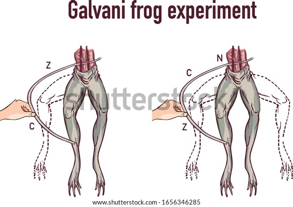
Galvani Experiment Frog Legs Vector Illustration Stock Vector Royalty Free 1656346285 from image.shutterstock.com Use the scissors to continue the incision up to the midline all the way through. Also known as the middle phalanx, the short pastern bone sits on top of the articulating joint of the pedal bone and underneath the long pastern bone. When you are exploring the frog anatomy you will find things that you might recognize, like the frog's heart, brain, lungs and intestines. The wall is simply that part of the hoof that is visible when the horse is standing. But on the inside there are many similarities among human, bird, and bat forearms. Malformations included missing limbs, missing digits, extra limbs, partial limbs. The best tool for the job is a scalpel, where you scrap the top of the head, right between the eyes. To do this, make transverse (horizontal) cuts just inside the arms and legs, and connect them with a lateral cut up the belly.
Malformations were found in 8 species of frogs and toads.
The pedal bone is the only bone of these three that is completely inside the actual hoof, while the pastern bones serve to connect the hoof to the rest of the leg. Both the dorsal and ventral roots unite immediately after coming out of the neural canal through intervertebral foramen. Each of the small bones forming the fingers. Clean hoof and paddock daily. Malformed frogs have been found throughout minnesota (fig. Dorsal root has a ganglion of nerve cells. The postcaval vein receives blood from the kidneys by 5 to 6 pairs of renal veins and a pair of gonadial veins (spermatic in male and ovarian in female) from gonads. Only the bottom portion of this bone extends as far as the hoof capsule. A collection of small bones makes up a frog's digits, or its fingers and toes. Pastern bone is the fetlock joint and above that the cannon bone of the lower leg. Most of the time, a frog has five toes on its back legs and four toes on its front legs. Lay the frog on its back, spread out its limbs, and pin them to the tray. It connects the tarsus with the first phalanges of the digits.
A collection of small bones makes up a frog's digits, or its fingers and toes. Pastern bone is the fetlock joint and above that the cannon bone of the lower leg. Lay the frog on its back, spread out its limbs, and pin them to the tray. Place the frog knee just beneath the transducer hook. They have a short backbone (spine), with a large hip bone to support their powerful leg muscles.
Comparison Between The Skeletons Of Frogs Humans from photos.demandstudios.com A frog has two scapulae, or shoulder blades, and clavicles, or collarbones, that are shaped a lot like the same bones in a person's body. These are the astragalus and the calcaneus. A third division of the frog's leg consists of two elongated anklebones, or tarsals. Anaerobic bacterial infection of the frog and commissure groove. For northern leopard frogs ( rana pipiens ), the species most commonly found in minnesota, 6.5 percent of 13,763 frogs collected were malformed. In indian frog, rana tigrina usually 9 pairs of spinal nerves are found which arise from the spinal cord by two roots, a dorsal or sensory root and a ventral or motor root. The postcaval vein receives blood from the kidneys by 5 to 6 pairs of renal veins and a pair of gonadial veins (spermatic in male and ovarian in female) from gonads. Pastern bone is the fetlock joint and above that the cannon bone of the lower leg.
Attaching the muscle to the transducer 1.
The development of a beak has led to evolution of a specially adapted. The frog's skeletal and muscular systems consist of its framework of bones and joints, to which nearly all the voluntary muscles of the body are attached. Voluntary muscles, which are those over which the frog has control, occur in pairs of flexors and extensors. Use the scissors to continue the incision up to the midline all the way through. Human, bird, and bat bone comparisonfrom the outside human arms, bird wings, and bats wings look very different. Severe cases may require packing with medication and padding the hoof. The best tool for the job is a scalpel, where you scrap the top of the head, right between the eyes. It connects the tarsus with the first phalanges of the digits. Dorsal root has a ganglion of nerve cells. Continue the cut up the center of the frog's body with scissors, being careful to cut through the skin only. These are the astragalus and the calcaneus. Lift the frog's skin with forceps between the rear legs. Each of the small bones forming the fingers.
Frogs have long, strong back legs, with extra joints so they can fold up close to the body leg bones diagram. Moisten the leg with ringers so you don't lose function from drying out.


0 Komentar